Bioherb -
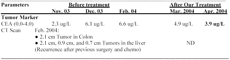
Colon Cancer Recurrence
54 years old patient with Colon Cancer Recurrence after surgery and Chemotherapy
Jan. 2004: Patient was told to go home as no treatment can work
| ( ) indicates normal range ND = Not Done | |
| Note: | |
| Feb. 2004: | Started our Cancer Treatment program |
| July 2004: | Doing great with fantastic health |
| Aug 30, 2004: | Patient visited an Oncologist and was suggested 4 doses of combined chemotherapy and cure. |
| Sep 2004: | Patient agreed to go for chemotherapy and our treatment was stopped. Unfortunately not 4 but 16 doses of chemotherapy did not work and patient died. |
71 years old patient with Colon Cancer spread to Liver
Patient received palliative chemotherapy FOLFIRI Protocol Oct. 2003 to March 2004
This protocol includes Irinotecan, Leucovorin and 5FU
Summary before our treatment: After chemotherapy patient was still weak and frail. Tumor Marker CEA reduced significantly from 150 in June 2003 to 7.8 in March 2004 but re-lapsed and increased to 19 in July 2004. At this point patient was too unwell to stand another chemo.
Patient started our treatment program in September 2004.
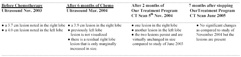
Note:
Chemotherapy was significant in reducing Tumor Marker but did not improve patient's health in general. Patient was sick and unwell even months after stopping chemotherapy.
Patient took our treatment only for 3 months (Sep. to Nov. 2004) and there was a great improvement in patient's quality of life and general health. Patient was in good health until January 2006 and by the end of Jan. 06 we heard that there was another re-lapse of Tumor Marker CEA.
Only those who know about Cancer treatment would appreciate the success of a treatment program that help a last stage cancer patient to live for 14 months with good health and quality. Some anti-Cancer drugs are approved based on claims that it prolongs the life of a dying patient by a few weeks. If that is the case then our treatment program is one of the best and yet the safest.
45 years old patient with history of Thyroid Cancer
Recovery with our Cancer Treatment Program and Radiation Therapy
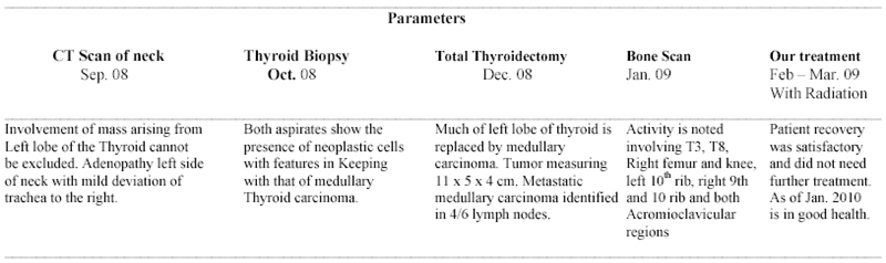
Summary:
Radiation therapy that was given after the first CT scan and biopsy report confirmation did not work.
Total Thyroidectomy was done in Dec. 08 and histopathology report indicated Medullary Carcinoma of thyroid involving lymph nodes.
Bone Scan of Jan. 08 was a bad news but not a surprise. Cancer spread to other parts of the body.
At this point patient came to us with pain, discomfort, and loss of normal voice (cancer effecting vocal cord).
First Feb. 2009 patient started our treatment and besides it in March 09, received 5 weeks of Radiation Therapy. Our treatment was stopped by the end of March.
As of Jan. 2010 patient is in good health.
74 years old patient with Left Renal Cell Carcinoma spread to Vertebra
| Summary before our treatment | |
| Jun 24, 2004: | CT Scan |
| July 2004: | Patient is anemic with low hemoglobin, abnormalities in blood tests, low chloride and sodium and high urea and creatinine |
| July 9, 2004: | MRI report confirms the CT Scan findings. Keeping in mind the age and fragile health no treatment (Surgery, Radiation or Chemotherapy) was recommended. Patient was put on high doses of Morphine |
| After our treatment | |
| Sep 7, 2004: | Started our Cancer Treatment Program |
| Oct 5, 2004: | Stopped Morphine completely and recovering very well. |
| Oct 2004 – June 2005: | Continued with our treatment with great success and recovery. |
84 years old patient with Lung Cancer
| Summary before our treatment | |
| Mar 30, 2004: | CT scan shows 2 cm mass in keeping with a primary malignancy on the left lower lobe. |
| May 27, 2004: | Some calcified lymph nodes are evident in the right paratracheal area and in the right hilum. There are 6 calcified nodules and 3 non-calcified demonstrated in the right lung which could be due to earlier old granulomatous infection. |
| Feb 9, 2005: | A mass lesion seen at the left lung base. The findings are suspicious for lung Cancer with suspected 4R Lymphadenopathy |
| Feb 16, 2005: | CT scan, left lower lobe of the lung, the mass measures 2.4 x 2.5 cm |
| Mar 3, 2005: | Doctors suspect it could certainly be "Bronchogenic Malignancy but she clearly would not tolerate Chemotherapy. Radiation might be possible but the overall effect on her survival would likely be negligible. Therapeutic options are severely limited and it would be appropriate to simply leave well enough alone." |
| After our treatment | |
| Sep 22, 2005: | Within a month of our treatment the patient's health improved drastically and continued the treatment for 4 months then stopped. Lived a normal life till 2008 and died in 2008 due to a fall that caused brain injury. |